Fig. 8.

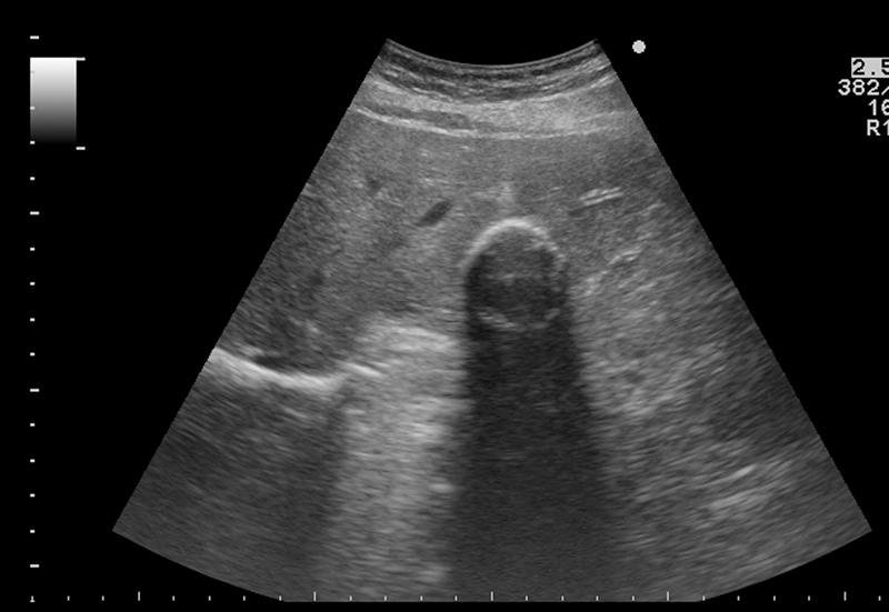
Appearance of a stage CE5 cyst. Ultrasound image of a CE5 cyst with the calcified rim clearly seen, together with a posterior acoustic shadowing.

Appearance of a stage CE5 cyst. Ultrasound image of a CE5 cyst with the calcified rim clearly seen, together with a posterior acoustic shadowing.