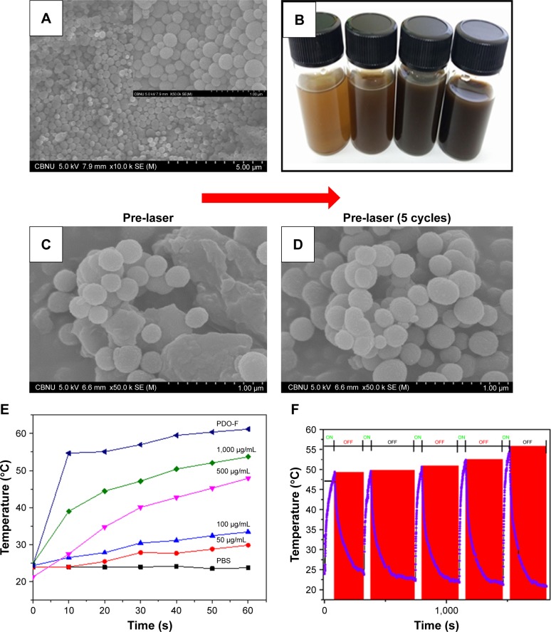
Figure 4.
Characterization of PD NPs and photothermal stability testing.
Notes: (A) SEM image of PD NPs (inset – the magnified image of A). (B) A vial containing an aqueous suspension of various concentrations of PD (50, 100, 500, and 1,000 µg/mL). (C and D) Stability of PD NPs before and after five cycles of laser irradiation (2 W/cm2; 808 nm at 60 seconds per cycle). (E) Effect of concentration on the photothermal output of PD NPs at 60 seconds irradiation (808 nm; 2 W/cm2). (F) High precision thermal cycle by nonstop laser on–off switching on PDO composite fiber. The blue and red zones as indicated represent “on” and “off” modes, respectively. The sample was irradiated with 808 nm laser at the power of 2 W/cm2 for 60 seconds (“on”) followed by natural cooling for 300 seconds (“off”).
Abbreviations: PD NPs, polydopamine nanospheres; PDO, polydioxanone; SEM, scanning electron microscopy.

