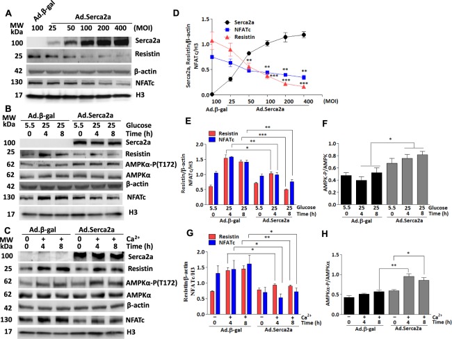
Figure 5.
Serca2a overexpression suppresses high glucose induced-resistin and NFATc expressions and enhanced AMPK activation. H9c2 cells were infected with increasing multiplication of infection (MOI) of Ad.Serca2a. The expression of Serca2a, resistin and NFATc (nuclear) was analyzed by western blotting (A) and densitometry quantifications were determined (D). **p < 0.01 and ***p < 0.001 vs baseline Ad.βgal. Serca2a-overexpressing H9c2 cells (MOI:50) were treated with high glucose concentration (25 mM vs. 5.5 mM) for the indicated times. The expression of resistin, nuclear NFATc and phosphorylation of AMPKα were analyzed by western blotting (B) and densitometry quantifications were obtained (E,F). Serca2a-overexpressing H9c2 cells were treated with 4 mM Ca2+ for the indicated times and the expression of resistin, nuclear NFATc, and phosphorylation of AMPKα were analyzed by western blotting (C) and densitometry quantification (G,H). The phosphorylation of AMPK status is presented as phospho-AMPK/AMPK ratio. β-actin was used as an internal control. The data are mean ± SEM of at least three experiments in triplicates. *p < 0.01, **p < 0.01 and ***p < 0.001 Ad.Serca2a vs Ad.β-gal at the indicated times.

