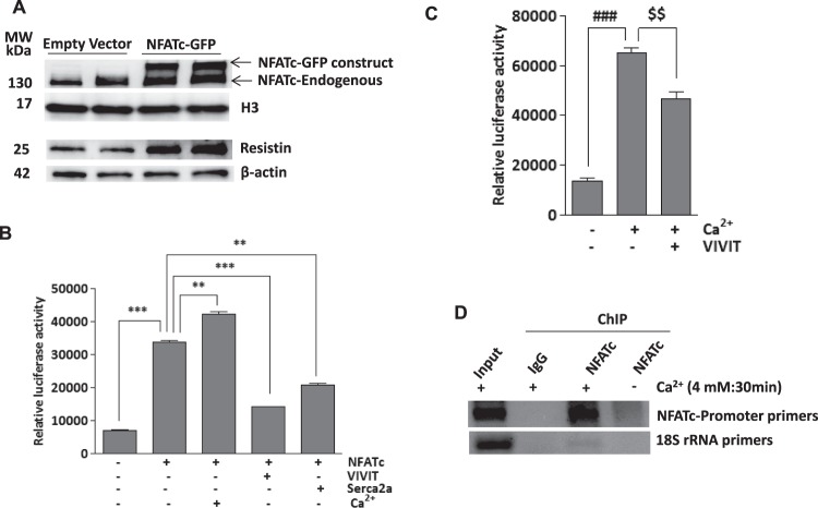
Figure 7.
NFATc induces resistin expression and activates resistin promoter activity- H9c2 cells were transiently transfected with Ad.NFATc-GFP or Ad.empty vectors for 48hrs and nuclear NFATc and resistin protein expression was measured by western blotting with H3 or β-actin used as loading controls, respectively (A). Myocytes were transduced with Ad.NFATc and resistin promoter-mediated luciferase activity was measured in the presence of VIVIT, Serca2a or Ca2+ (4 mM) (B). Myocytes transduced with Ad.VIVIT were stimulated with 4 mM Ca2+ and resistin promoter-mediated luciferase activity was measured as indicated (C). Chromatin immunoprecipitation (ChIP) assay was performed to determine binding of NFATc to resistin transcription loci in the NFATc overexpressing or control H9c2 cells stimulated with Ca2+ (4 mM) for 30 min. The agarose gel picture of the PCR products shows relative binding of NFATc to a specific region of resistin promoter, precipitated with either NFATc or IgG antibody. 18S rRNA was used as control (D). **p < 0.01 NFATc vs Ca2+ and Serca2a; ***p < 0.001 Cont vs NFATc, NFATc vs VIVIT (B). $$p < 0.001 Ca2+ vs VIVIT ###p < 0.01 vs Ca2+.

