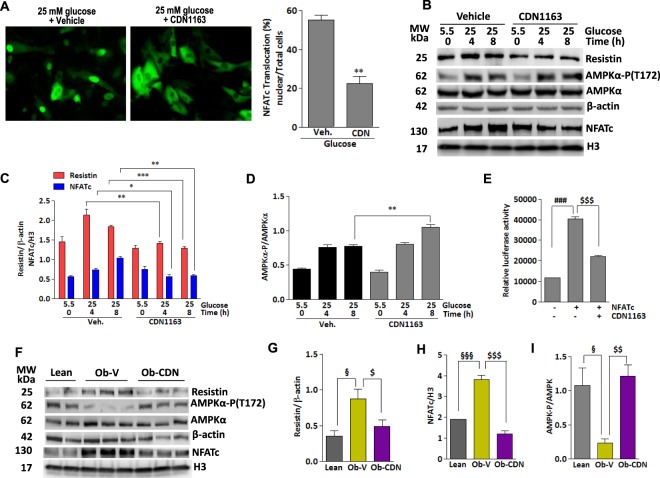
Figure 8.
Pharmacological activation of Serca2a with small molecule CDN1163 inactivates NFATc and reduces resistin expression. (A) Representative fluorescence microscopic images of H9c2 cells transfected with NFATc-GFP for 24 hours and then incubated with CDN1163 (10 μM) or vehicle for an additional 24 hours. Cells were then stimulated with high glucose for 4 hours and nuclear import of NFATc-GFP was visualized and quantified in more than 5 different images per condition (A). **p < 0.01 CDN1163 vs Veh. (B) H9c2 cells were treated with CDN1163 and stimulated with high glucose concentration (25 mM vs. 5.5 mM) for the indicated times. The expression of resistin, nuclear NFATc proteins, and phosphorylation of AMPKα were analyzed by western blotting (B) with densitometry quantifications determined (C,D). Resistin promoter-mediated luciferase activity was measured in H9c2 cells overexpressing NFATc then treated with CDN1163 for 24 hours (E). Ob/ob mice were injected with CDN1163 (Ob-CDN) or vehicle (Ob-V) for 4 weeks and heart tissues were analyzed for the expression of resistin, nuclear NFATc and phosphorylation of AMPKα by western blotting (F) with densitometry quantifications shown (G–I, respectively) with H3 or β-actin used as corresponding internal controls. The phosphorylation of AMPK status is presented as phospho-AMPK/AMPK ratio. The data are mean ± SEM of three experiments from 4–5 animals. *p < 0.05, **p < 0.01 and ***p < 0.001 veh vs CDN1163 at the indicated times (C,D); ###p < 0.001 Cont vs NFATc; $$$p < 0.001 NFATc vs CDN1163; §p < 0.05 and §§§p < 0.001 lean vs Ob-V; $p < 0.01, $$p < 0.01 and $$$p < 0.001 Ob-V vs CDN1163.

