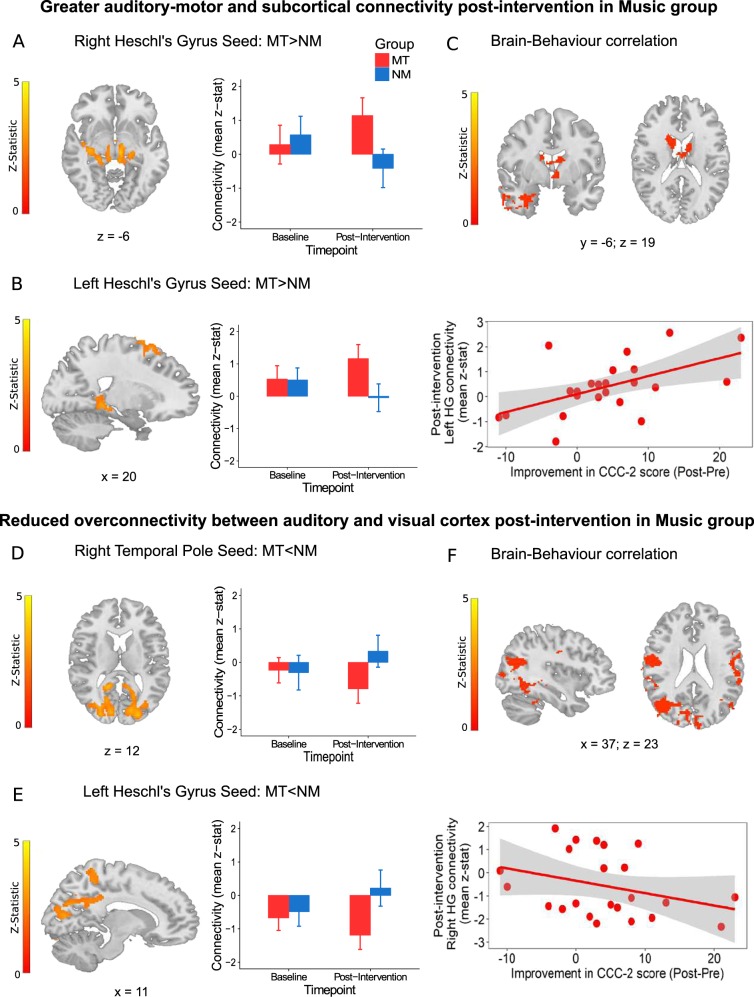
Fig. 3. Brain functional connectivity outcomes and correlation with behavioural improvement.
The top panel shows regions of increased resting-state functional connectivity (RSFC) post-intervention in the Music (MT) vs. Non-music (NM) groups between a Right Heschl’s gyrus seed and subcortical regions such as the hippocampus and thalamus (z = 3.94, P < .0001) and b left Heschl’s gyrus seed and fronto-motor regions (z = 3.16, P < .0001). c Connectivity between auditory and subcortical thalamic and striatal regions post-intervention is directly related to improvements in communication measured using the change in CCC-2 composite score in MT (z = 3.57, P < .0001). The bottom panel shows regions of decreased RSFC post-intervention in MT vs. NM groups between d right temporal pole seed and occipital regions (z = 4.01, P < .00001) and e left Heschl’s gyrus seed and bilateral calcarine and cuneus regions (z = 3.39, P < .00001). f Connectivity between auditory and visual sensory cortices post-intervention is inversely related to improvements in communication measured using the change in CCC-2 composite score in MT (z = 3.64, P < .001). MT is shown in red and NM in blue. Errors bars represent standard error (SE). All brain images are presented in radiological convention and coordinates are in MNI space

