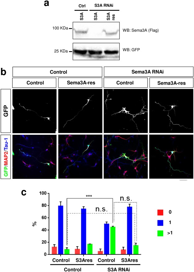
Figure 2.
Knockdown of Sema3A induces the formation of supernumerary axons. (a) HEK 293 T cells were transfected with vectors for FLAG-Sema3A (S3A) or RNAi-resistant FLAG-Sema3A-res (S3Ares) and an shRNA directed against Sema3A (S3A RNAi) or pcDNA6.2-GW/EmGFP-miR (Ctrl). The expression of Sema3A and GFP was analyzed by Western blot (WB) using an anti-FLAG antibody. The molecular weight is indicated in kDa. (b) Hippocampal neurons from E18 rat embryos were transfected with vectors for GFP (green), an shRNA against Sema3A (Sema3A RNAi) or pcDNA6.2-GW/EmGFP-miR (control), and a vector for RNAi-resistant FLAG-Sema3A-res (Sema3A-res) or pBK-CMV (control) as indicated. Neurons were analyzed at 3 d.i.v. by staining with an anti-MAP2 (red, dendrites) and the Tau-1 antibody (blue, axon). Representative images of transfected neurons are shown. The scale bar is 25 μm. (c) The percentage of unpolarized neurons without an axon (0, red), polarized neurons with a single axon (1, blue) and neurons with multiple axons (>1, green) is shown (Student’s t-test and two-way ANOVA; n = 3, independent experiments with >150 neurons per experiment; values are means ± s.e.m, ***p < 0.001 compared to control as indicated; n.s., not significant).

