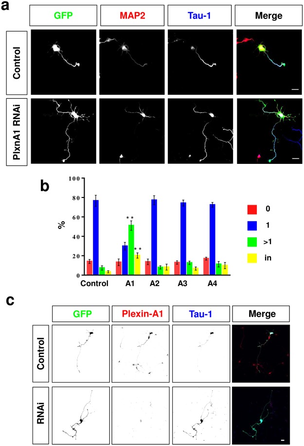
Figure 4.
Knockdown of Plexin-A1 induces the formation of supernumerary axons. (a,b) Hippocampal neurons from E18 rat embryos were transfected with vectors for GFP (green) and shRNAs against Plexin-A1, -A2. -A3, or -A4 or pSUPER (control) and analyzed at 3 d.i.v. by staining with an anti-MAP2 (red, dendrites) and the Tau-1 antibody (blue, axon). Representative images of transfected neurons are shown. The scale bar is 25 μm. (b) The percentage of unpolarized neurons without an axon (0, red), polarized neurons with a single axon (1, blue), neurons with multiple axons (>1, green) and neurons with neurites that are positive for both axonal and dendritic markers (in, yellow) is shown (Student’s t-test and two-way ANOVA; n = 3, independent experiments with >150 neurons per experiment; values are means ± s.e.m, **p < 0.01 compared to control). (c) Hippocampal neurons from E18 rat embryos were transfected with vectors for GFP (green) and an shRNA against Plexin-A1 (RNAi) or pSUPER (control) and analyzed at 3 d.i.v. by staining with an anti-Plexin-A1 (red) and the Tau-1 antibody (blue, axon). Representative images of transfected neurons are shown. The scale bar is 25 μm.

