Figure 1.
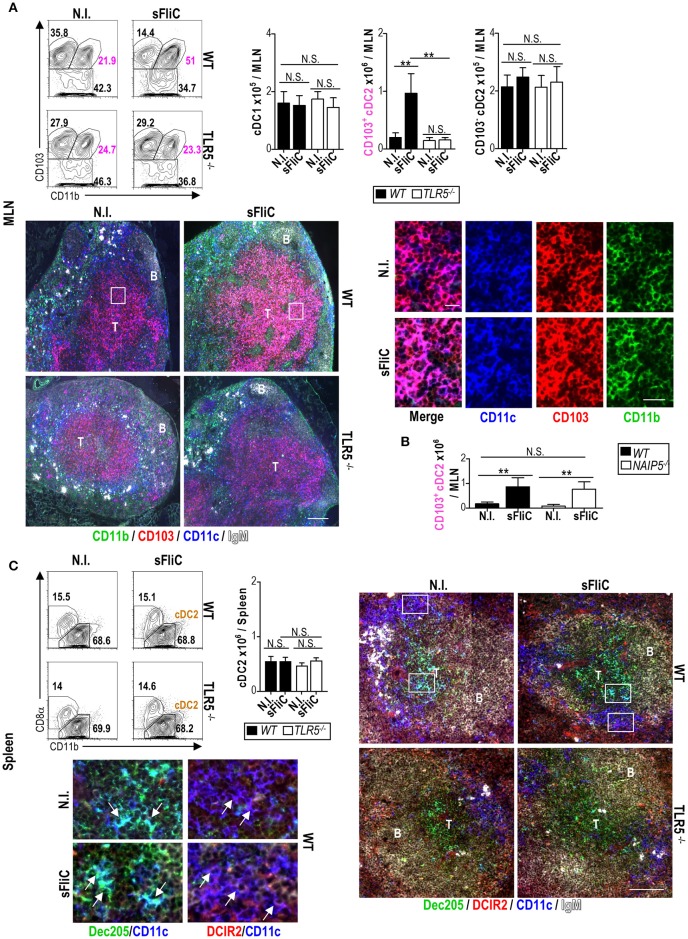
Mucosal CD103+cDC2 respond to sFliC immunization in a TLR5-dependent manner. Wild-type (WT) or TLR5−/− mice were immunized i.p. with sFliC and cDCs (Lin−MHC-IIhiCD11chi) were evaluated 24 h later, alongside non-immunized (N.I.) mice. (A) MLN representative flow cytometry plots (including percentages) of cDC1 (Lin−MHC-IIhiCD11chiCD11b−CD103+), CD103+cDC2s (Lin−MHC-IIhiCD11chiCD11b+CD103+) and CD103−cDC2 (Lin−MHC-IIhiCD11chiCD103−) are shown with adjacent graphs of absolute numbers. Representative photomicrographs of MLN sections stained for CD11c; blue, CD103; red, CD11b; green, and IgM; white (scale bar = 200 μm) are shown (top right). Zoom-in insets (white boxes) show single staining and a merge of CD11c, CD103, and CD11b (scale bar = 20 μm). T, T zone; B, B zone. (B) WT or NAIP5−/− mice were immunized i.p., with sFliC and absolute numbers of MLN CD103+cDC2s were evaluated 24 h later, alongside non-immunized (N.I.) mice. (C) Wild-type (WT) or TLR5−/− mice were immunized i.p. with sFliC and splenic cDCs (Lin−MHC-IIhiCD11chi) were evaluated 24 h later, alongside non-immunized (N.I.) mice. Representative flow cytometry plots (including percentages) of cDC2s (Lin−MHC-IIhiCD11chiCD11b+) are shown with adjacent graphs of absolute numbers. Representative photomicrographs of spleen sections stained for CD11c; blue, Dec205; green, DCIR2; red, and IgM; white (scale bar = 100 μm). Zoom-in insets (white boxes) show the differential location of cDC1s (Dec205+) in the T zone and cDC2s (DCIR2+) in the bridging channels. Data shown as mean+s.d. of 4 mice and are representative of 3 independent experiments. **P < 0.001, by two-way analysis of variance (ANOVA) N.S., not significant.

