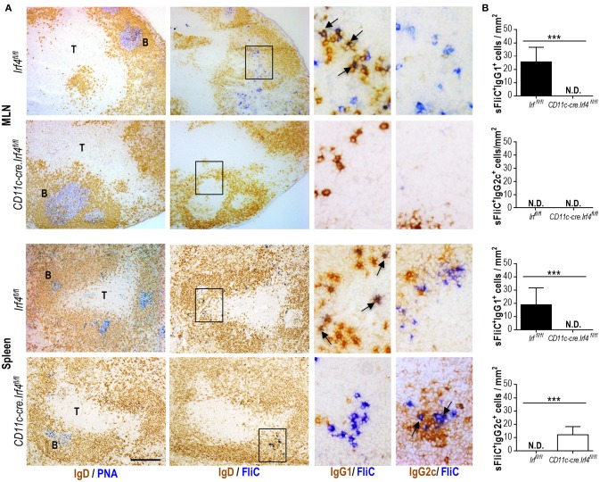
Figure 4.
Switching to IgG1 is abrogated in the CD11c-cre.Irf4fl/fl mice. Irf4fl/fl or Cd11c-cre.Irf4fl/fl mice were sFliC-immunized for 7 days. (A) Representative photomicrographs of serial sections from MLN and spleen stained for: PNA-binding cells (blue) and IgD-expressing cells (brown) (first column) or sFliC-binding cells (blue) and IgD-expressing cells (brown) (second column), scale bar = 200 μm. The third and fourth columns show zoom-in insets (black-boxed areas) stained to detect sFliC-binding cells and IgG1 and IgG2c respectively (scale bar = 50 μm). T, T zone; B, B zone. (B) Quantification of sFliC+IgG1+ cells and sFliC+IgG2c+ cells in the MLN and spleen. A total of 10 random fields were evaluated per slide. Data shown as mean + s.d. (n = 8 mice pooled from two independent experiments). ***P < 0.0001, by Mann-Whitney. N.D. non-detected.

