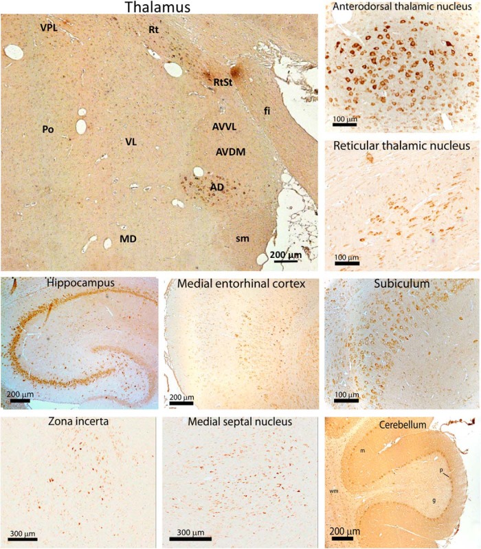
Figure 2.
CACHD1 protein expression in adult rat brain. Immunoreactive protein was detected using rabbit anti-CACHD1 with peroxidase anti-rabbit secondary antibody and DAB staining (brown). AD, Anterodorsal thalamic nucleus; AVDM, anteroventral thalamic nucleus (dorsomedial); AVVL, anteroventral thalamic nucleus (ventrolateral); fi, fimbria; MD, mediodorsal thalamic nucleus; Po, posterior thalamic nucleus; sm, strai medullaris; Rt, reticular thalamus nucleus; RtSt, reticular VL, ventrolateral thalamic nucleus; VPL, ventroposterior lateral thalamus; g, granule cell layer; m, molecular layer; p, Purkinje cell; wm, white matter. Figure 2 is supported by expression profiling of CACHD1 and different voltage-gated calcium channel subunit mRNA in human tissue (Figure 2-1).

