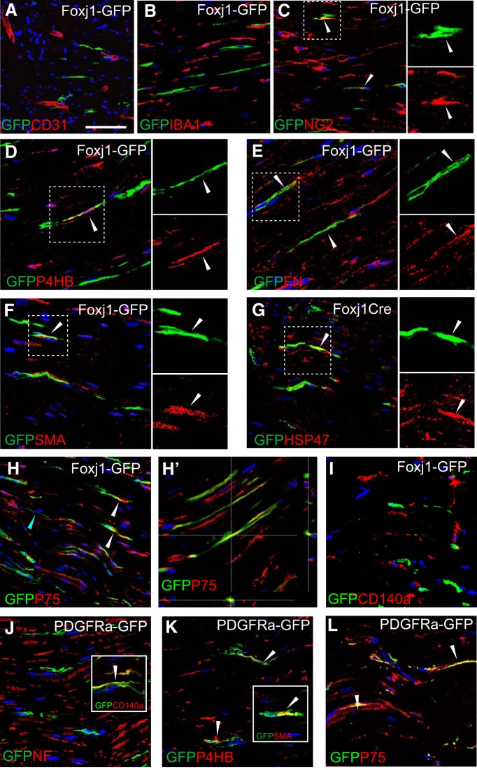
Figure 2.
Foxj1-GFP-labeled cells express fibroblast markers but are of a distinct population from PDGFRa-GFP labeled cells in peripheral nerves. Immunofluorescence characterization of normal sciatic nerves (SN) from Foxj1-GFP mice treated with tamoxifen. Double immunostaining for GFP and various markers was performed on longitudinal sections. Colocalized staining is marked with arrowheads. Overlaid images show that Foxj1 labeled cells in SN are not associated with endothelial cells labeled by CD31 (A), nor do they express the macrophage marker IBA1 (B). A proportion of GFP+ cells are labeled with NG2 (C) and proteins usually expressed by fibroblasts such as fibronectin (FN, D), P4HB (E), SMA (F), and HSP47 (G). In D–G, the split channel views of the boxed area by dotted lines are shown in separate images. Foxj1-GFP-labeled cells also expressed P75NTRs (H and confocal orthogonal view in H′). GFP-labeled cells do not coexpress PDGFRa (CD140a, I). PDGFRa fate-mapping GFP reporter mice were characterized by double immunostaining. PDGFRa-GFP labeled cells are not associated with neurofilament-labeled axons (J). The GFP+ cells are confirmed expressing CD140a (inset in J) and are double labeled with P4HB and SMA (K and inset in K). PDGFRa-GFP-labeled cells are also detected for P75NTR (L). Scale bar indicates 50 μm for all images.

