Figure 3.
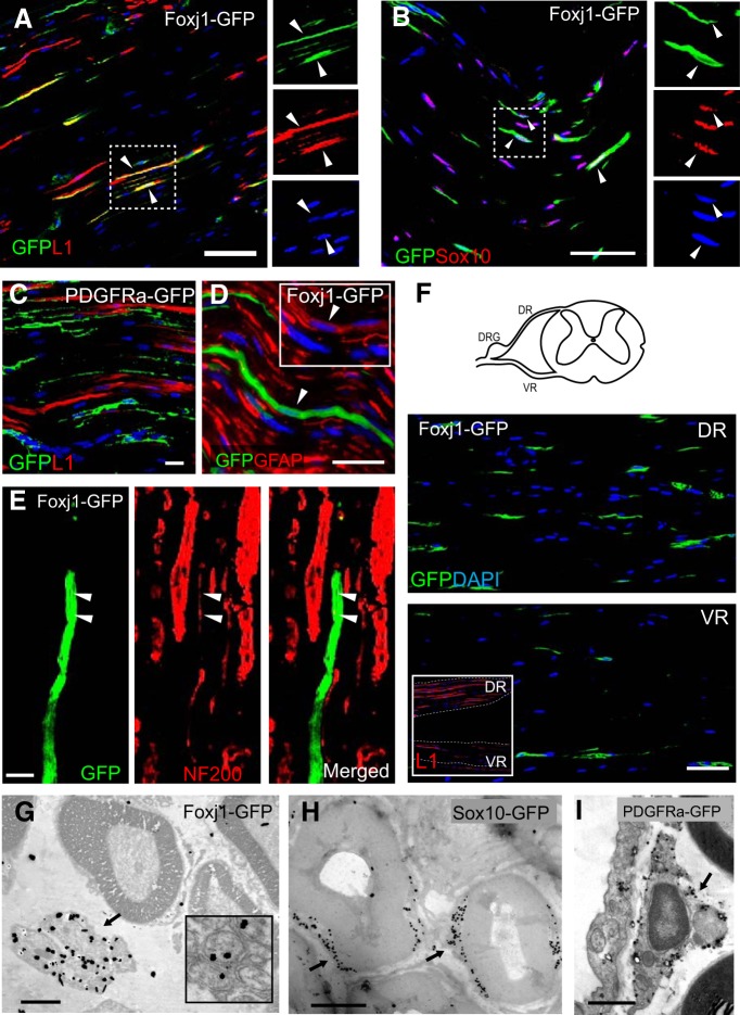
Foxj1 labels nonmyelinating SCs. Images in A–F illustrate merged double immunofluorescence staining on longitudinal sections of sciatic nerve (SN) from adult Foxj1-GFP mice treated with tamoxifen. Most GFP-expressing cells coexpress the L1 cell adhesion molecule, a marker of nonmyelinating SCs in peripheral nerves (A). GFP-expressing cells can also express Sox10, a neural crest-related transcription factor (B). The single channel images of the same area in A and B marked by dotted line are shown in image sets on the right side of the main images. PDGFRa-labeled cells (PDGFRa-GFP) in sciatic nerves do not coexpress L1 (C). Low levels of GFAP are detected in Foxj1-GFP-expressing cells (D, inset). Costaining for GFP and neurofilament reveals a close association between GFP and small-diameter axons (arrowheads) (E). Dorsal and ventral roots (DR and VR, respectively) from lumbar spinal cord were stained for GFP, showing greater numbers of Foxj1-GFP+ cells in dorsal root (F) and this was correlated with the proportion of L1 immunoreactivity in corresponding locations (inset in F). G–I are images from pre-embedding immunogold electron microscopy against GFP on SN from Foxj1 and Sox10 fate-mapping mice. Silver-enhanced gold particles were mainly detected on “Remak” bundles in Foxj1-GFP samples (G), whereas the majority of gold particles in Sox10 fate-mapping mice are deposited in SC cytoplasm around the myelinated axons but excluded from compact myelin sheaths (H). PDGFRa labeling identifies endoneurial fibroblast like cells (I). Scale bars: A–E, 25 μm; F, 50 μm; G, 2 μm; H, I, 1 μm.

