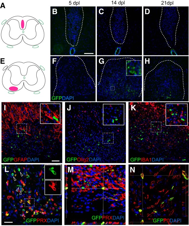
Figure 5.
Foxj1-labeled cells give rise to remyelinating SCs in demyelinating lesions in spinal cord white matter. Demyelination lesions were induced in mice Foxj1-GFP-expressing mice by direct injection of lysolecithin in either dorsal or ventral funiculi as shown in A and E, with corresponding images from GFP immunostaining for 5, 14, and 21 dpl. Few GFP-expressing cells have been detected in dorsal lesions (B–D), whereas a large number of GFP+ cells are found in ventral lesions from 14 dpl (F–H). Double immunostaining indicates that Foxj1-GFP cells do not express the astrocyte marker GFAP (I), the oligodendrocyte lineage marker Olig2 (J), nor the microglial marker IBA1 (K). L shows that considerable numbers of GFP-expressing cells coexpress the myelinating SC marker PRX (arrows). The box area is shown as split channels (L1 and L2) and merged in (L3). M, Orthogonal view of confocal images of double immunostaining confirming the colocalization of GFP and PRX in ventral lesions at 14 dpl. The SC identity of GFP+ cells in lesion is verified by colocalization with myelin P0, as illustrated by orthogonal confocal view in N. Insets in the images show magnified area in lesions in respective images marked with dotted outlines, for either double staining (I–K) or single colors (G, L). Scale bars in B indicate 100 μm and apply to C, D, and F–H. Scale bar in I indicates 50 μm and applies to J and K. In L–N, the scale bars indicate 25 μm.

