Figure 6.
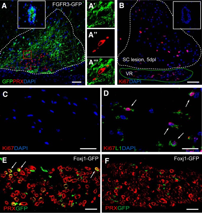
SC remyelination from Foxj1-GFP-labeled cells in spinal cord lesions are not derived from ependymal cells, but likely originate from peripheral nerves. A, Demyelinated lesion at 21 dpl in ventral spinal cord white matter of a FGFR3-GFP reporter mouse treated with tamoxifen. Image shows overlaid double immunostaining for GFP and PRX, with the dotted line marking the boundaries of the lesion. GFP is expressed by ependymal cells lining the central canal of the spinal cord (inset). A magnified area with in the red box in the lesion in A showing separate and overlaid channels indicating nonoverlapping expression of GFP and PRX is shown in A′, A″, and A‴). B illustrates a ventral spinal cord lesion (white dotted line) at 5 dpl immunostained for the proliferation marker Ki67. Ki67+ cells are found in the adjacent ventral roots (VR, green dotted line), but not in ependymal cells in central canal (inset). There are no Ki67+ cells in the contralateral VR (C). The Ki67+ nucleus are found in L1+ cells in the VR of ventral spinal cord lesion side (D). E and F illustrate Foxj1-GFP cells colabeled with PRX in the VR of spinal cord lesion side at 14 dpl, but none in the VR of contralateral side. Scale bars indicate 100 μm in A, 50 μm in B, and 20 μm in C–F.

