Figure 7.
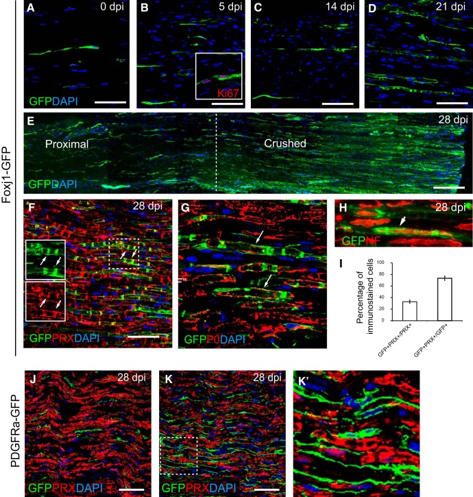
Foxj1-GFP-labeled cells become repair SCs following sciatic nerve injury. Sciatic nerve crush lesions were created in adult mice expressing reporter genes and samples were analyzed by immunofluorescence staining. A–D show an area of sciatic nerve in longitudinal section immunostained to reveal Foxj1-GFP-labeled cells in normal (A) and crushed nerve at 5, 14, and 21 dpi (B–D, respectively). Inset in B illustrates GFP+ cells expressing Ki67. E is a montage combined from a series of overlapping images spanning the proximal and crushed site at 28 dpi. Doubling immunostaining shows that, at 28 dpl, GFP colocalizes with the mature myelinating SC marker PRX (F). The dotted outlined area is shown in single channels as in insets in F, with examples of colocalization marked with arrows. GFP labels the myelinated axons marked by myelin P0, which appears only localized in the cytoplasm rather than compact myelin (G). GFP-labeled cells enclose the axons (arrows) marked by neurofilament (NF) staining (H). Approximately 30% of remyelinated axons in the crush area are labeled with GFP, but >70% of GFP immunoreactivity colocalizes with PRX (mean ± SE, n = 3; I). J and K show GFP and PRX double immunostaining in control (uninjured nerve) and crushed sciatic nerve from PDGFRa-GFP animals at 28 dpi. The magnified boxed area in K is shown in K′ which shows that there is no colocalization between GFP and PRX. Scale bars indicate 50 μm for all images.

