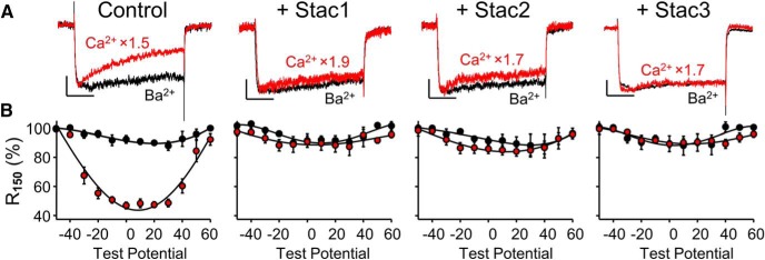
Figure 1.
Stac proteins suppress Ca2+-dependent inactivation of l-type Ca2+ currents in neonatal rat hippocampal neurons. Representative whole-cell Ca2+ (red, vertically scaled by indicated factors) and Ba2+ (black) currents at +10 mV (A) and fraction of peak current remaining 150 ms after the peak (R150) as a function of test potential (B) are shown for either control neurons or neurons transfected with Stac1-, Stac2-, or Stac3-YFP (left to right, respectively). l-type currents were isolated by pharmacological blockade of non-l-type currents (see Materials and Methods). Numbers of cells (Ca2+, Ba2+): (9,4), (7,6), (6,3), and (6,4) for control, Stac1-, Stac2-, and Stac3-transfected cells, respectively. Here and in subsequent figures, the error bars indicate ± SEM. Scale bars in A: 10 pA/pF (unscaled Ba2+ currents, vertical), 50 ms (horizontal).

