. 2018 Sep 3;32(5):1071–1080. doi: 10.21873/invivo.11348

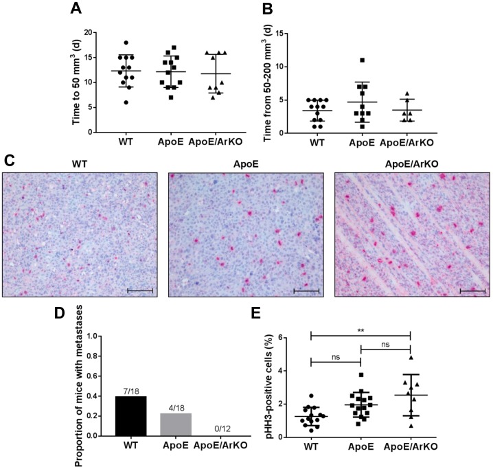
Figure 2. Tumours from ApoE−/−/ArKO mice were significantly more proliferative than tumours from wild-type (WT) mice. A: Analysis of the lagphase of tumour growth as time in days (d) to reach 50 mm3 in wild-type, ApoE−/− and ApoE−/−/ArKO mice. Wild-type and ApoE: n=12;ApoE/ArKO: n=9. B: Analysis of the exponential phase of tumour growth as time in days to quadruple in volume. Wild-type: n=12; ApoE: n=10;ApoE/ArKO: n=6. C: Representative immunohistochemical staining for the mitotic marker phosphohistone H3 (pHH3) in tumours from wild-type,ApoE−/− and ApoE−/−/ArKO mice. Scale bars represent 100 μm. D: Proportion of wild-type, ApoE−/− and ApoE−/−/ArKO mice with internalmetastases. E: Analysis of tumour cell proliferation from wild-type, ApoE−/− and ApoE−/−/ArKO mice according to the percentage of pHH3-positivecells. Wild-type: n=14; ApoE: n=15; ApoE/ArKO: n=9. **Significantly different at p<0.01, ns: not significant. Data are expressed as individualdata points and mean±SD.

Figure 2