Figure 1.
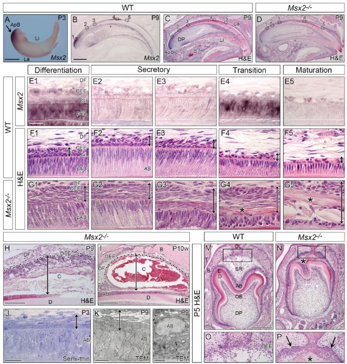
Msx2 expression in WT and morphological phenotypes in mutant incisors and molars. Genotypes and stages are indicated. (A–L) Incisal edge side to right. (A) Whole-mount in situ hybridization of the lower incisor showing intense Msx2 expression in the apical bud (ApB) region (arrow). (B–I) Section in situ hybridization of Msx2 and hematoxylin and eosin (H&E)–stained sagittally sectioned upper incisors. (E–G) Magnified views of boxed areas in B to D. Msx2 is expressed in the outer enamel epithelium (OEE), stratum intermedium (SI), and ameloblasts (AB) at the differentiation (E1) and transition (E4) stages but downregulated at the secretory (E2–3) and maturation (E5) stages. Mutant ameloblasts polarize normally at the differentiation and early secretory stages (G1–2) but depolarize abnormally afterward (G3–5). A stratified squamous epithelium forms ectopically in the mutant enamel organ (double-headed arrow in G1–5). An eosinophilic keratin-like structure is also observed (asterisk in G4–5). (H, I) An odontogenic cyst forms in the mutant enamel organ. Double-headed arrow indicates cyst thickness and dotted line demarcates epithelial-mesenchymal border. (J–L) Semi-thin analysis and transmission electron microscopy confirm the stratified squamous epithelium (double-headed arrow) and depolarized ameloblasts. (M–P) Frontally sectioned lower first molars. (O, P) Magnified views of boxed area in M and N. A stratified squamous epithelium ectopically forms and an eosinophilic keratin-like structure exists in the mutant enamel organ (asterisk and arrows in P). B, bone; C, cyst; D, dentin; DF, dental follicle; DL, dental lamina; DP, dental pulp; E, enamel; La, labial side; Li, lingual side; LI, lower incisor; OB, odontoblast; SR, stellate reticulum; T, tongue; WT, wild-type. Bars: 500 µm in A, 500 µm in B for B–D, 25 µm in E1 for E–G, 100 µm in H, 250 µm in I, 25 µm in J, 15 µm in K, 10 µm in L, and 250 µm in M for M and N.

