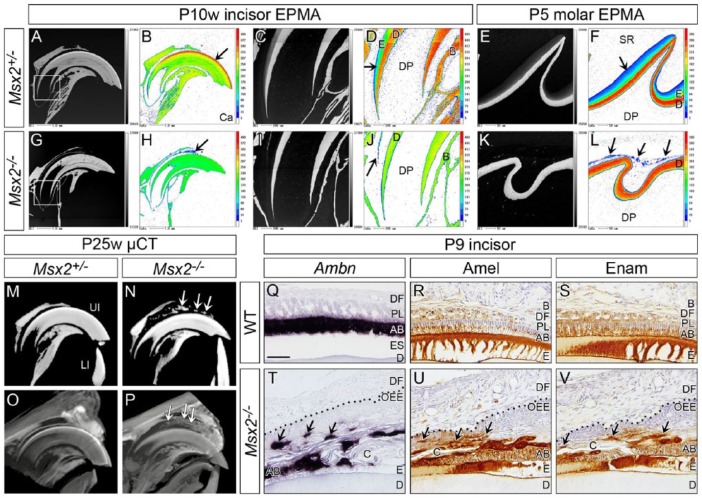
Figure 4.
Ectopic amorphous mineralized structures are observed in the mutant enamel organ. Genotypes and stages are indicated. (A–L) Sagittal views obtained by electron probe microanalyzer (EPMA). (B, D, F, H, J, L) Calcium (Ca). (A–D, G–J) Upper incisors (incisal edge side to right). (C, D, I, J) Magnified views of boxed area in A and G. Compared to control, almost no enamel is on dentin and ectopic mineralized structures are observed between the incisor and surrounding bone in Msx2−/− (arrow in H, J). (E, F, K, L) Upper first molars (distal side to right). Similarly, almost no enamel forms and ectopic mineralized structures exist within the mutant enamel organ (arrows in L). Note that color scale is different depending on the magnification. In magnified views of controls (D, F), the color intensity of enamel is lower than that of dentin because enamel is immature and not fully mineralized at this stage. (M–P) Sagittal views of micro–computed tomography (µ-CT) analysis of upper incisors (incisal edge side to right). (M, N) Ectopic mineralized structures are detected between the incisor and surrounding bone in Msx2−/− (arrows in N). (O, P) A contrast stain using phosphotungstic acid reveals that such mineralized structures exist within the odontogenic cyst (arrows in P; see also Appendix Fig. 16). (Q–V) In situ hybridization of Ameloblastin (Ambn) and immunohistochemistry for Amelogenin (Amel) and Enamelin (Enam) of sagittally sectioned upper incisors (incisal edge side to right). Dotted line in T–V demarcates epithelial-mesenchymal border. Enamel protein-producing ameloblast-like cells localize to the cystic wall (arrows in T, U, V). AB, ameloblast; B, bone; C, cyst; D, dentin; DF, dental follicle; DP, dental pulp; E, enamel; ES, enamel space; LI, lower incisor; OEE, outer enamel epithelium; PL, papillary layer; SR, stellate reticulum; UI, upper incisor. Bar: 50 µm in Q for Q–V.

