Fig. 2.

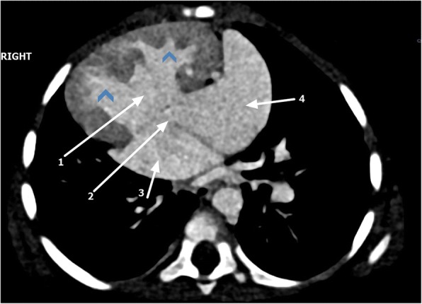
Cardiac computed tomography axial view showing dextroposition of the heart, ventricular septal defect (1), atrial septal defect (2), double right atrium (3, 4), and transposed ventricles (arrowheads)

Cardiac computed tomography axial view showing dextroposition of the heart, ventricular septal defect (1), atrial septal defect (2), double right atrium (3, 4), and transposed ventricles (arrowheads)