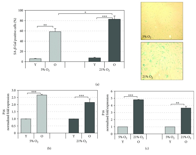
Figure 2.
Markers of cellular aging. (a) Quantitative analysis of positive β-galactosidase-stained cells in HDF during cellular aging under 5% and 21% O2 culture conditions. The percentage of blue cells observed in old cells (right panel) under a standard light microscope was calculated. Error bars represent ± SEM. (b) p16 mRNA modulation during aging in old (O) HDF with respect to young (Y) ones cultured under normoxic (21% O2) and hypoxic (5% O2) conditions; (c) p16 mRNA expression in cells cultured under 21% O2 with respect to 5% O2 (right panel). The gene was normalized vs GAPDH/β-actin/SDHA, and the results are reported as normalized fold expression. Error bars represent ± SD ∗ p < 0.05; ∗∗ p < 0.01; ∗∗∗ p < 0.001.

