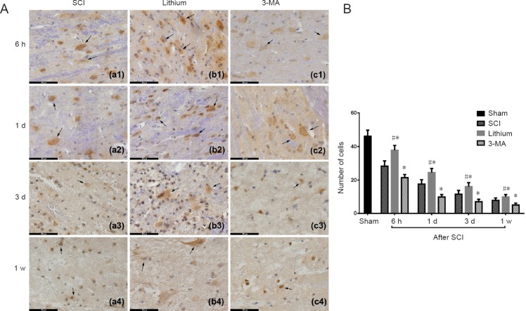
Figure 3.
Immunohistochemical staining and counting of neurons (NeuN+ cells) in the injured rat spinal cord at different time points.
(A) Neurons were more numerous in the lithium group than in the other groups at 6 hours, 1 and 3 days, and 1 week. Arrows point to neurons. Scale bars: 50 μm. (B) Neurons in each experimental group were significantly decreased at 1 and 3 days and 1 week after SCI compared with the previous time point. More neurons survived in the lithium group than in the SCI group, while more neurons survived in the SCI group than in the 3-MA group. All data are expressed as the mean ± SD (n = 3; one-way analysis of variance followed by the least significant difference post hoc test). *P < 0.05, vs. SCI group; #P < 0.05, vs. 3-MA group. SCI: Spinal cord injury; 3-MA: 3-methyladenine; h: hours; d: day(s); w: week.

