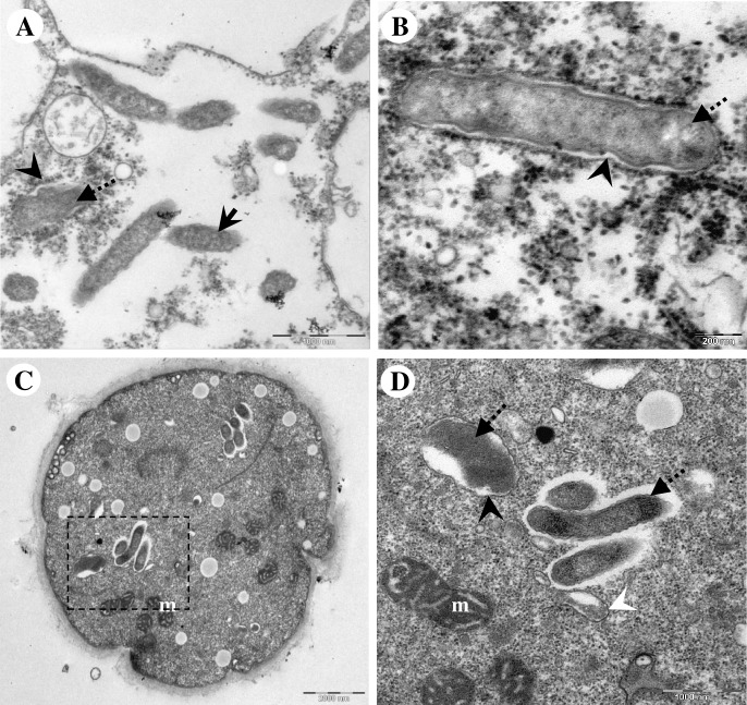
Fig 6. Transmission electron microscopic images demonstrating unknown membranous structure which selectively surrounded the morphologically altered endocytobionts.
(A) Early form of membranous structure appending to a morphologically altered endocytobiont. Note the presence of intact endocytobionts in the surrounding. (B) Higher magnification showing a formed membranous vacuole which contained a morphologically altered endocytobiont. (C) Unknown membraneous vacuole observed inside a cyst of Acanthamoeba sp. HTH136. (D) Higher magnification of the square with dotted-line in (C). Note the membrane-bound endocytobiont appears disintegrated, and the vacuolar membrane is not entirely closed. Nearby are three endocytobiotic bacteria; the surface of these cells appeared slightly rough, however, they were not encircled by vacuole. An autophagic isolation membrane which engulfed cytoplasmic cargo is seen. Indicators = unknown membranous structure: ‘black arrow-head’; morphologically altered endocytobionts: ‘dotted-arrows’, and morphologically intact endocytobionts: ‘solid-arrows’, autophagic isolation membrane: ‘white arrow-head’, and mitochondria: m.

