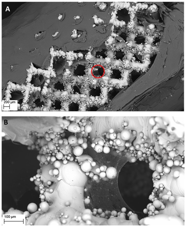
Fig 2. Visualization of cellulose covering (A) and ingrowing (B) the pores of Ti6Al7Nb implant.
The area marked by a red circle in (A) is visualized with higher magnification in (B). The cellulose seen in (A) was intentionally mechanically disrupted (as seen in the middle of the picture) to uncover the underlying implant’s struts. Magn. 51x and 318, respectively. Zeiss EVO MA SEM Microscope. Please also refer to S3 Fig to see more Ti6Al7Nb implants with partially removed cellulose.

