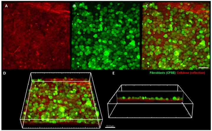
Fig 4. Fibroblasts colonizing bacterial cellulose membrane.
Cellulose is visualized using laser reflection (shown in red) (A), while fibroblasts are fluorescently labeled (shown in green) (B). Merged channels are presented in (C) (view from the top), (D) and (E) (side views). The imaging was performed on a Leica SP8 confocal microscope.

