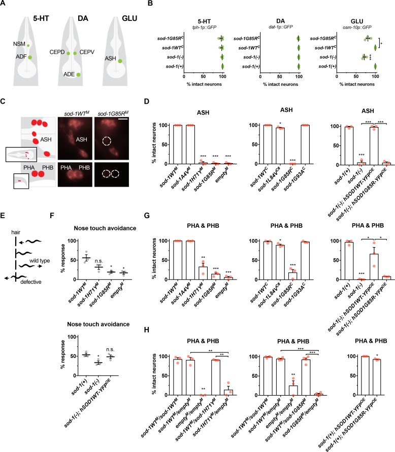
Fig 5. sod-1 loss of function drives oxidative stress induced glutamatergic neuron degeneration in single-copy/knock-in ALS sod-1 animals.
(A) Illustration of glutamatergic (osm-10p::GFP), serotonergic (tph-1p::GFP) and dopaminergic (dat-1p::GFP) neurons examined to assess neurodegeneration after paraquat-induced oxidative stress. (B) Serotonergic (5-HT) and dopaminergic (DA) neurons were unaffected in all genotypes examined. However, glutamatergic neurons (GLU) were lost after paraquat-induced oxidative stress in sod-1(-) and sod-1G85RC animals, compared to sod-1(+) and sod-1WTC controls, respectively. sod-1(+) designates the unedited wild type gene at the endogenous locus; this is the same allele present in standard N2 strain. (C) Left: illustration of six glutamatergic sensory head neurons (top) and two tail neurons (bottom) labelled with lipophilic retrograde dye DiD. Images show right side of animal; similar neurons on left side were also scored. Middle: representative images of sod-1WTM animals with intact glutamatergic sensory neurons in the head (top) and tail (bottom) labelled with DiD after paraquat treatment. Right: representative images of sod-1G85RM animals; ASH (top), PHA and PHB (bottom) neurons fail to take up dye only after paraquat treatment, indicating either process degeneration/retraction or neuronal loss. Scale bar represents 10 μm. (D) Percent intact ASH neurons in ALS model animals after paraquat treatment, compared to appropriate sod-1WTM, sod-1WTC, hSOD1WT-YFPOE or sod-1(+) wild type controls. Degeneration in ASH neurons was increased in sod-1H71YM, sod-1G85RM, sod-1L84VC, sod-1G85RC and hSOD1G85R-YFPOE animals, as well as in animals lacking sod-1 function (emptyM controls and sod-1(-)). sod-1L84VC indicated with #. sod-1(+) is the standard N2 strain. In all figure panels, results of three independent trials are shown. Error bars indicate ±SEM. N > 25 for all genotypes in all panels. Two-tailed t-test: * P < 0.05; ** P < 0.01; *** P < 0.001. (E) Illustration of the nose-touch avoidance assay. Wild type animals initiate backward locomotion after their nose contacts a hair; animals that cross over the hair or fail to reverse are scored as defective. ASH sensory neurons play an important role in this behavior [39]. Animals were treated with 2.5 mM paraquat for 4 hours before the nose touch response assay. After 4 hours of paraquat treatment, nose touch response was modestly impaired in wild type controls (57% response), even though 98% of ASH neurons were intact (3 trials, 35 animals total). After the same 4 hour treatment, nose touch response was severely impaired in sod-1G85RM animals (28% response), while 49% of ASH neurons were intact (3 trials, 35 animals total). (F) Top: sod-1G85RM animals and emptyM control animals lacking sod-1 function were defective in response to nose-touch after paraquat treatment compared to sod-1WTM control animals. Bottom: Neuronal expression of the human SOD1WT-YFP in sod-1(-) animals restored nose-touch response. sod-1(+) is standard N2 strain. (G) Percent intact glutamatergic PHA and PHB neurons in ALS model animals after paraquat treatment, compared to appropriate sod-1WTM, sod-1WTC, or sod-1(+) wild type control animals. Degeneration was increased in PHA and PHB neurons of sod-1H71YM, sod-1G85RM and sod-1G85RC animals compared to appropriate wild type controls. Loss of sod-1 function in emptyM control and sod-1(-) animals also led to PHA and PHB degeneration, compared to sod-1WTM and sod-1(+). sod-1(+) is the standard N2 strain. Neuronal overexpression of human SOD1WT-YFP, but not human SOD1G85R-YFP, partially restored dye-uptake in sod-1(-) animals. (H) To examine the consequences of altering gene dosage and assess recessive/dominance of single-copy/knock-in ALS alleles, homozygous ALS sod-1 animals and controls were crossed to homozygous sod-1WTM or emptyM males carrying a GFP-expressing transgene, and cross-progeny were tested for DiD dye-uptake after paraquat treatment. emptyM/emptyM cross-progeny had defects after paraquat treatment, compared to sod-1WTM/WTM cross-progeny, while sod-1WTM/emptyM animals had intact glutamatergic neurons. Heterozygous sod-1H71YM/emptyM and sod-1G85RM/emptyM animals were defective, compared to heterozygous sod-1WTM/emptyM animals. By contrast, sod-1WTM/sod-1H71YM or sod-1WTM/sod-1G85RM animals had intact glutamatergic neurons after paraquat treatment. Additionally, animals overexpressing the human hSOD1G85R-YFP protein in the sod-1(+) background had low penetrance dye-filling defects. sod-1(+) designates the unedited wild type gene at the endogenous locus; this is the same allele present in standard N2 strain.

