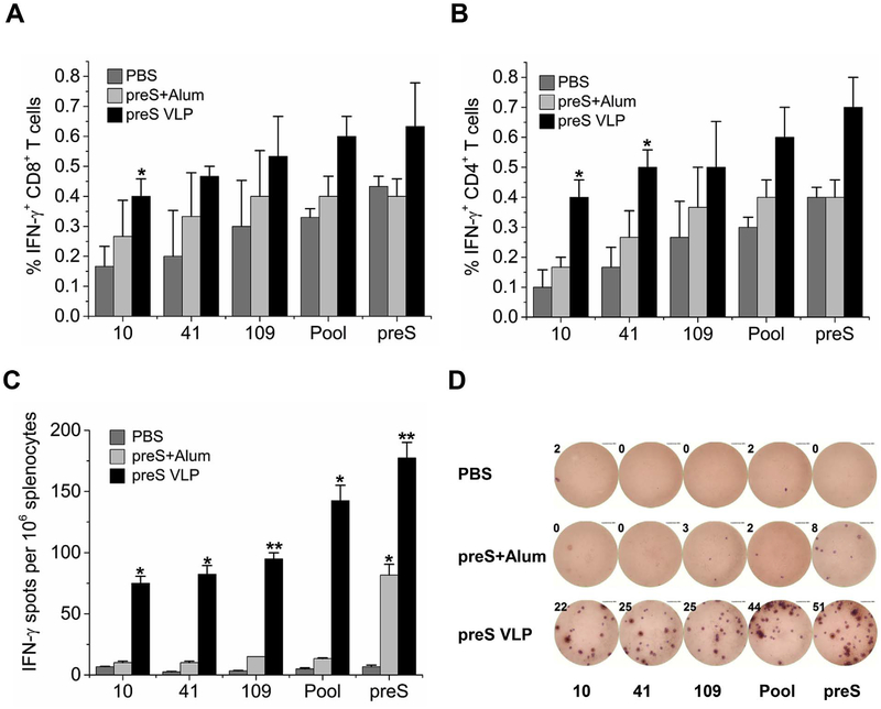
Fig. 4. preS V LP induces stronger T cell responses than recombinant preS vaccination.
Balb/c mice were immunized intramuscularly with preS VLP (n = 6), recombinant preS protein (n = 6), or PBS (n = 6). 30 days postimmunization, splenocytes were isolated and analyzed for CD8, CD4, and IFN-γ expression by flow cytometry (A–B). CD8+ T cells (A) or CD4+ T cells (B) were gated, and IFN-γ-producing cells are presented as the percent average from each group. (C) Splenocytes were isolated and analyzed for IFN-γ expression using an IFN-γ ELISPOT assay. ELISPOT experiments were performed in triplicate wells per condition. (D) Representative images of ELISPOT from each group are shown. All values are presented as the average from each group, and error bars indicate ± SEM. Pool, indicates the mixture of peptide 10, 41 and 109, and preS indicates recombinant preS protein.

