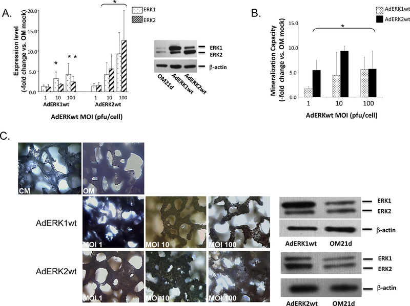
Figure 8: Overexpression of ERK1/2 increases ASC mineralization.
Panel A - Overexpression of ERK1 or ERK2 by OM-induced ASC monolayers transduced by recombinant ERK1 or ERK2 adenoviruses (AdERK1wt or AdERK2wt) at the indicated MOIs (pfu/cell) was confirmed after 21 days through western blotting and compared with OM mock controls (Expression, -fold change vs. OM mock). ERK1/2 overexpression is shown in a representative Western blot. Panel B- Mineralization by transduced ASCs was quantified after 21 days of OM induction and compared with controls (Mineralization Capacity, -fold change vs. OM mock). Panel C - 3D hydroxyapatite-coated PLGA scaffolds seeded with AdERK1wt- or AdERK2wt-transduced ASCs and cultured in OM for 21 days are shown after von Kossa staining to detect mineralization. Staining within a scaffold layer is shown compared with controls maintained in CM or OM. A representative Western blot showing isoform-specific overexpression of ERK1/2 following transduction with AdERK1wt or AdERK2wt at MOI 10 is shown on the right. For Panels A and B, * indicates a significance difference (p<0.05; t-test) between groups.

