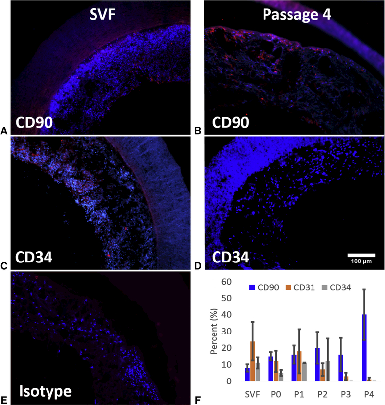
Figure 1:
Representative IFC images depicting positive staining of cells for CD90 (A and B) and CD34 (C and D). Initial SVF cells stained positively for both CD90 (A) and CD34 (C), while positive staining for CD90 (B) increased and positive staining for CD34 (D) decreased at passage 4. E) Isotype control demonstrating no non-specific immunopositive cells. F) IFC analysis of percentage of adherent cells positive for cell markers CD90 (mesenchymal stem cell marker), CD31 (endothelial marker), and CD34 (endothelial progenitor marker) seeded onto PEUU scaffolds with cells from fresh SVF out to passage 0 through 4 and given a 48 hour dynamic culture period.

