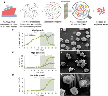
Fig. 1. Coculturing of E. huxleyi with Sulfitobacter D7 isolate exhibits distinct phases of pathogenicity.

(A) A scheme describing the origin of the CAM bacterial consortium and isolation of Sulfitobacter D7 from an E. huxleyi bloom in the North Atlantic. (B to D) A detailed time course of E. huxleyi 379 monocultures (gray line) and during coculturing with Sulfitobacter D7 (green line). The following parameters were assessed: (B) algal growth, (C) algal cell death, and (D) bacterial growth. No bacterial growth was observed in control cultures. The green background represents the presence of a pungent scent in cocultures. Algae-bacteria coculturing had distinct dynamics characterized by defined phases (1 to 3) of pathogenicity. (E to G) Scanning electron microscopy (SEM) images of (E) uninfected E. huxleyi 379 and (F and G) Sulfitobacter D7–infected E. huxleyi cells at phase 2 (scale bars, 2 μm). Arrows in (F) point to Sulfitobacter D7 attachment to E. huxleyi cells. Arrow in (G) points to a membrane blebbing–like feature. Results depicted in (B) to (D) represent average ± SD (n = 3). Error bars smaller than the symbol size are not shown. Statistical differences in (B) to (D) were tested using repeated-measures analysis of variance (ANOVA). P < 0.001 for the differences between control and cocultures.
