Fig. 1.

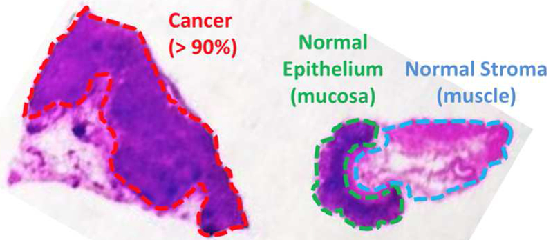
Histopathological assessment of a banked tissue example. This hematoxylin and eosin stain has been hand-labeled by a pathologist, marking three regions: gastric adenocarcinoma (cancer), epithelium (normal) and stroma (normal).

Histopathological assessment of a banked tissue example. This hematoxylin and eosin stain has been hand-labeled by a pathologist, marking three regions: gastric adenocarcinoma (cancer), epithelium (normal) and stroma (normal).