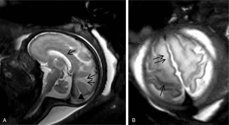
Figure 1.

Magnetic resonance imaging (MRI) of the fetal brain at 32 weeks gestation. Sagittal T2 turbo-spin echo (TSE) single-shot image shows the corpus callosum (arrow), the parieto-occipital (double arrows) and calcarine (arrowhead) sulcus [A, Axial T2 TSE single shot image discloses the central sulcus (arrow); B, the superior frontal gyrus (double arrows)].
