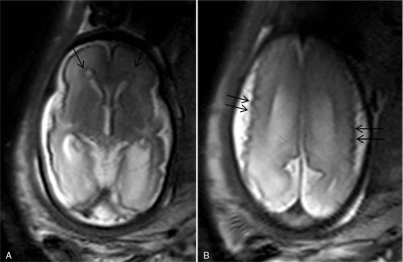
Figure 3.

A 33 weeks gestation patient was referred for magnetic resonance imaging (MRI) because a positive cytomegalovirus serology at the first pregnancy trimester, and cerebral fetal lesions viewed by sonography (not shown). Axial T2 turbo-spin echo (TSE) single-shot images shows microcephaly, micropolygyria (double arrows), and periventricular cavities (arrows) (A and B).
