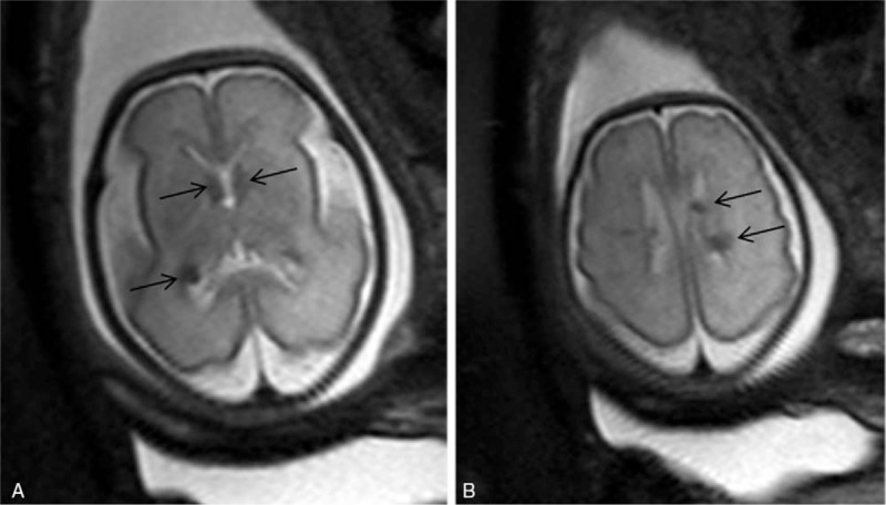
Figure 4.

Cardiac rhabdomyomas were diagnosed by sonography in a 28 weeks gestation suggesting tuberous sclerosis but the fetal brain sonogram was normal. Axial T2 turbo-spin echo (TSE) single-shot magnetic resonance (MR) images show round hypointense (arrows) images located at the ependyma and adjacent to the Monro foramen, confirming Tuberous Sclerosis with cerebral lesions (A and B). These findings were confirmed at autopsy.
