
Figure-3.
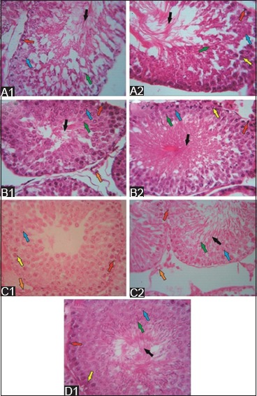
The structure of seminiferous tubules after secretome injection in the low- and high-dose groups (hematoxylin and eosin staining; 500×). 1 week after secretome injection, few spermatogonia (red arrow) and spermatozoa (black arrow) were observed in the low-dose group (A1), and few primary spermatocytes (blue arrow) were observed in the high-dose group (A2). The regenerative effects were observed in both groups 1 week after the second secretome injection. That is, the spermatogenic cells were complete. However, spermatozoa were denser in the seminiferous tubules (B2) of the high-dose group than in those of the low-dose group (B1). 1 week after the third secretome injection, low- and high-dose group (C1 and C2) showed complete spermatogenic cells. However, no spermatozoa were noted in the low-dose group. 1 week after the fourth secretome injection in the high-dose group (D1), the complete stage of spermatogenic cells and spermatozoa was observed in the lumen of the seminiferous tubules.