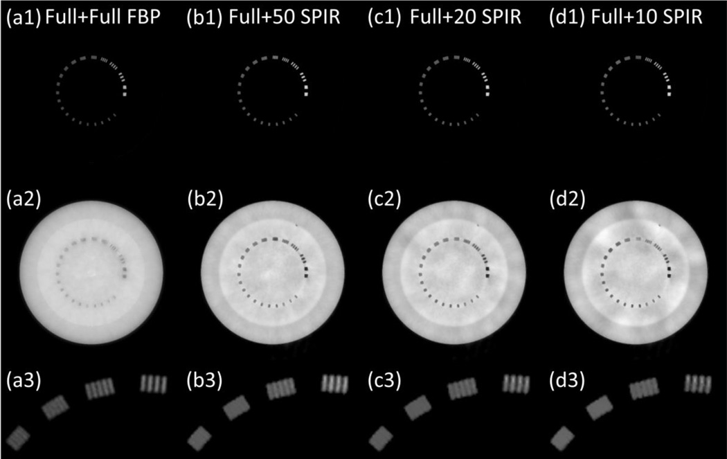
Figure 4.
The decomposed material images of line pair slice, based on the reconstruction images at low kVp and high kVp using (a) conventional two-full-scan FBP, SPIR-based DECT using one full scan and a second (b) 50-view, (c) 20-view and (d) 10-view scan. Row (1): “bone” images; row (2): “tissue” images; and row (3): magnified views of bone images in the ROI of dashed rectangle in Fig. 5(a). Display window: [0.1, 1.2].

