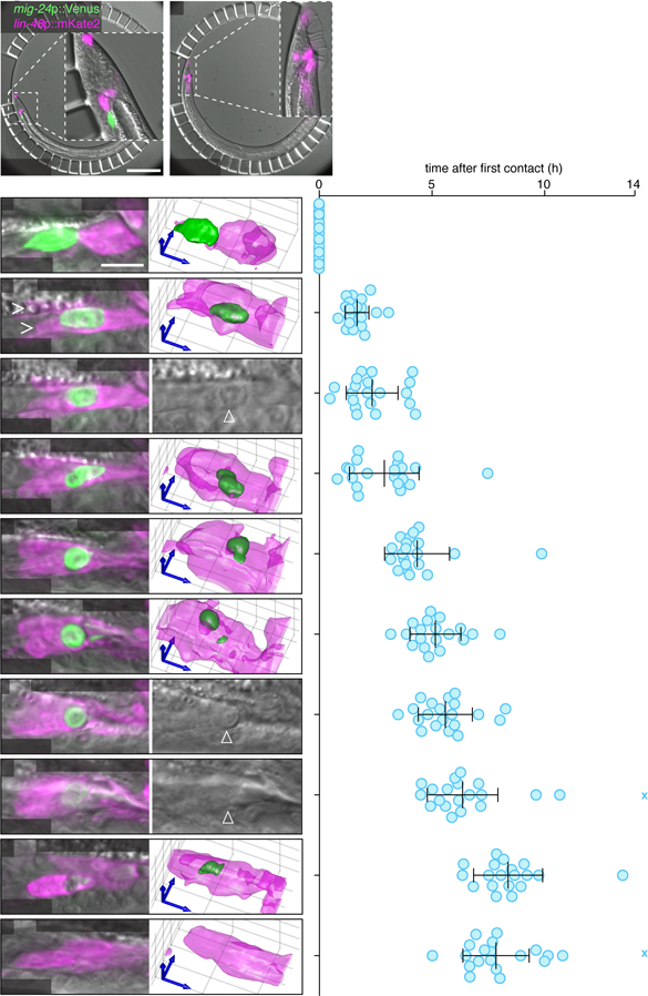
Figure 1. Linker cell death and degradation.

(A,B) C. elegans male immobilized in a microfluidic chamber (see (Keil et al., 2017)) at (A) first contact between linker cell (green) and engulfing U.l/rp cell (magenta, asterisk), and (B) after 10 hours. All males examined in all figures carry the male-producing him-5(e1490) or him-8(e1489) mutation. Scale bar, 100 µm. (C) Left: Maximum intensity projection showing linker cell at first contact with U.l/rp cells. Right: 3D rendering. Scale bar, 10 µm. (D-L) Stereotypical events during linker cell dismantling. Images as in (C), except that in (E,I,J) a single DIC slice is shown instead of 3D rendering. Right, time of events in individual animals (blue circles) after first contact. X, event did not occur during imaging. Bars, mean ± sd. Arrowhead, linker cell. (D) Linker cell between U.l/rp cells (caret) (E) Nuclear crenellation onset. (F) Competitive phagocytosis begins. (G) Linker cell rounding. (H) Linker cell corpse splits. (I) Onset of refractility. (J) End of refractility. (K) Small fragment disappears. (L) Large fragment disappears. See also Movies S1-S2, Figure S1, and Table S1.
