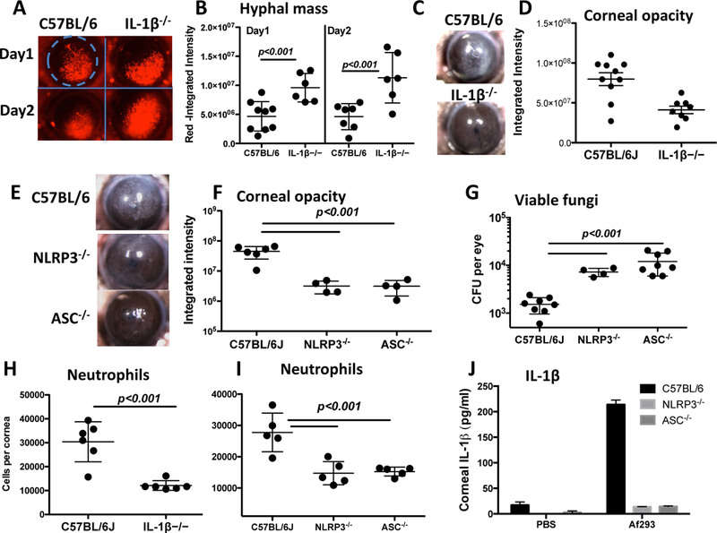
Figure 1. Impaired fungal killing in A. fumigatus infected IL-1β−/−, NLRP3−/− and ASC−/− mice.

A-D: Corneas of IL-1β−/− mice infected with A. fumigatus conidia and examined for hyphal growth (A, B), and for corneal opacification (C, D) as shown by representative images (A, C) (original magnification is x20). Hyphal mass and corneal opacity were quantified by image analysis (B, D). Data points represent individual corneas. E-G: Corneal opacification (E, F) and colony forming units (CFU, G) in infected NLRP3−/− and ASC−/− corneas. H, I: Neutrophil numbers in infected corneas following collagenase digestion, and total Ly6G+ cells per cornea was quantified by flow cytometry. J: Total IL-1β in homogenized corneas of NLRP3−/− and ASC−/− mice measured by ELISA. P values were calculated by 2 way ANOVA with Tukey post analysis (3 or more data sets) or by unpaired students t test (2 data sets). Data are representative of three repeat experiments.
