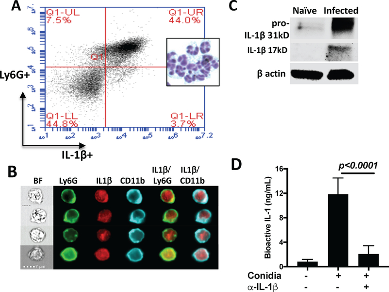
Figure 2. Neutrophils produce IL-1β in A. fumigatus corneal infection.

A: A. fumigatus infected corneas of C57BL/6 mice were collagenase digested 24h post infection, and Ly6G+ and intracellular IL-1β were quantified by flow cytometry. Inset shows polymorphonuclear cells from infected corneas after staining with Wrights-Giemsa. B: Representative neutrophils from infected corneas immunostained with Ly6G (NIMPR-14) and intracellular IL-1β examined by Imagestream analysis. C.Infected corneas were homogenized, run on SDS-PAGE, and IL-1β was detected by western blot. D. Homogenates of infected corneas were incubated with IL-1R reporter HEK cells, and total bioactive IL-1 was quantified. Data in A and C are from 5 corneas pooled together; Data in panel D are mean+/− SD from corneas from individual mice. All these experiments were repeated twice with similar results.
