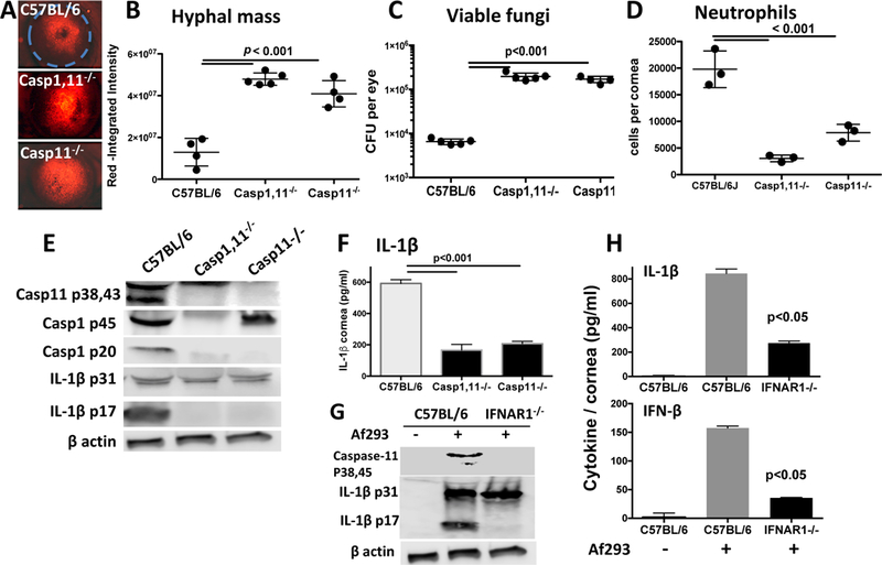
Figure 3. Impaired fungal killing and IL-1β cleavage in infected Caspase-11−/− mice.

A-D: Corneas of caspase-1−/− and caspase-1/11−/− mice 24h post-infection with A. fumigatus conidia. Representative RFP hyphae in infected corneas (A), and hyphal mass for each cornea was quantified by image analysis (B). Fungal viability was measured by CFU (C), and total Ly6G+ neutrophils per cornea were quantified by flow cytometry (D). Experiments were performed as described in the legend to Figure 1; data points represent individual corneas. E-H: Corneas from infected C57BL/6, caspase-1−/− and caspase-1/11−/− mice were homogenized and examined for pro- and cleaved forms of IL-1β (E), and secreted IL-1β (F). G,H. Corneas from C57BL/6 and IFNAR−/− mice were examined for pro- and cleaved IL-1β (G), and secreted IL-1β and IFN-β (H). production in infected corneas from control and IFNAR−/− mice (I). A: original magnification was x20; B-D: data points represent individual mice; E,G: Pooled corneas from three mice; F, H: mean+/−SD from 3–5 mice. P values were derived by ANOVA with a Tukey post-test.
