Figure 5.
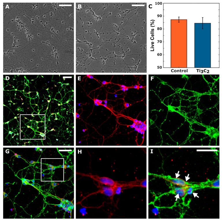
Biocompatibility of Ti3C2 MXene films. Representative phase images of primary cortical neurons cultured on (A) polystyrene and (B) 200 nm thick Ti3C2 films at 7 DIV. (C) Quantitative comparison shows no significant difference in neuronal survival on Ti3C2 or polystyrene (p > 0.6). (D–I) Representative multiphoton images of neurons cultured on Ti3C2 at 7 DIV stained for nuclei (blue), axons (red), and synapses (green). Outlined region in panel D is shown in panels E–G; the outlined region in panel G is shown in panels H and I, highlighting the network formation as confirmed by the presence of synapsin-positive puncta along the axons and cell bodies (arrows). Scale bars: 100 μm (panels A, B, and D), 50 μm (panel G), 25 μm (panel I).

