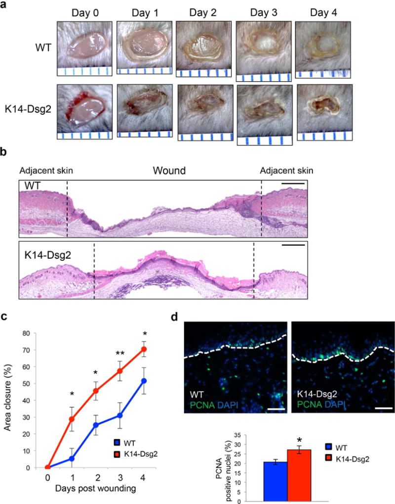
Figure 2. Dsg2 promotes cutaneous wound healing.

(a) Full-thickness wounds were inflicted on the dorsal skin of wild-type and K14-Dsg2 transgenic mice and photographed up to 4 days post-wounding. Space between blue ruler lines is 1 mm. (b) H&E staining of 4-day old wild-type and transgenic wounds. Dashed lines demarcate wound edge. Scale bar=100 μm (c) Wound area of non-epithelialized tissue was measured in ImageJ (mm2) and normalized to the area of fresh wounds. Data are expressed as average relative area of re-epithelialized tissue covering the initial wound (mm2) ± SEM. N=13 wild-type; 16 transgenic. (d) Representative immunofluorescence of wound-adjacent wild-type and transgenic mouse skin 4 days post wounding for PCNA (green) and DAPI (blue). Scale bars=50 μm.
