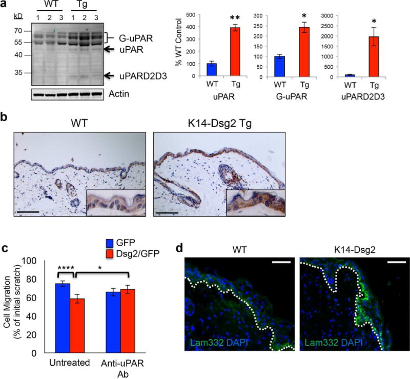
Figure 4. Dsg2 enhances epidermal and dermal uPAR in wounded tissues.

(a) Wild-type and K14-Dsg2 transgenic mouse epidermis (N=3) was immunoblotted for uPAR and actin. Signals were quantified, normalized to actin, and expressed as % of wild-type (right). G-uPAR: glycosylated uPAR; uPAR: non-glycosylated uPAR; uPARD2D3: D2D3 domains of uPAR. (b) Wild-type and K14-Dsg2 transgenic mice skins were immunostained for uPAR. (c) HaCaT+GFP or +Dsg2/GFP were treated with 1μg/mL anti-uPAR antibody and allowed to migrate for 24 h following scratch. Data are normalized to initial wound width of respective conditions and expressed as % distance remaining to be re-epithelialized. N=3. (d) Wild-type and K14-Dsg2 wounds (day 1) were immunostained for Laminin-332. Dashed line: regenerating basement membrane. Scale bars=50 μm.
