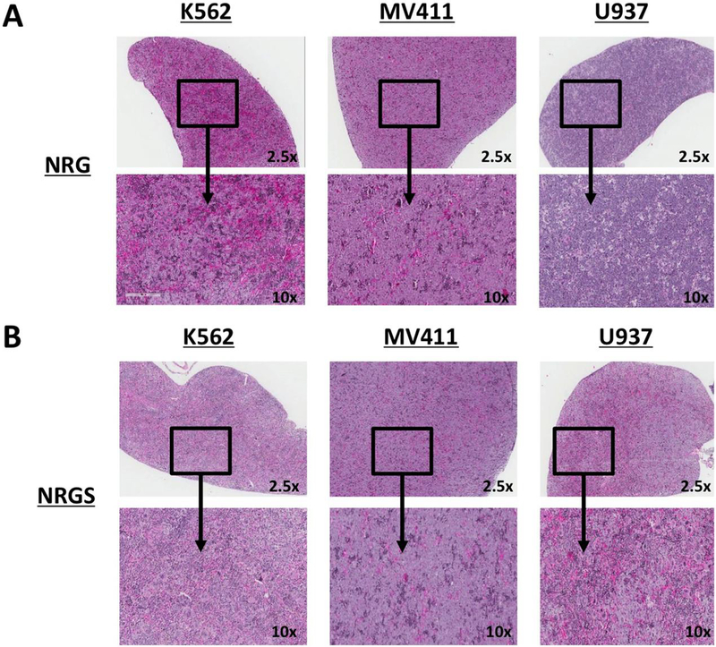
Figure 2. H&E staining of spleen sections from NRG and NRGS mice xenografted with established human cell lines.

(A) H&E stained sections of representative formalin fixed spleens spleens from NRG mice intravenously xenografted with 5 × 10^5 K562, MV411, or U937 cells per mouse suspended in 200ul of PBS. (B) Representative spleens from NRGS mice intravenously xenografted with 5 × 10^5 K562, MV411, or U937 cells per mouse suspended in 200ul of PBS.
