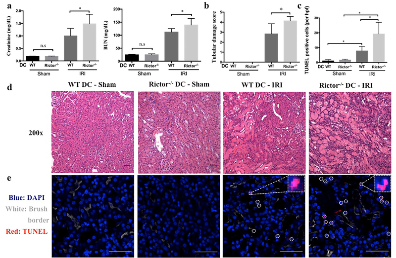
Figure 4|. Rictor deficiency in CD11c+ DC worsens acute kidney injury.
Age-matched male CD11cCre−/−Rictorf/f (WT DC) or CD11cCre+/+Rictorf/f (Rictor−/− DC) mice were challenged with sham operation or 20 min of bilateral renal ischemia and 24 hr reperfusion (n=3–10 mice per group). (a) Serum creatinine and BUN levels. (b-e) Representative appearances and quantitative analyses of hematoxylin and eosin- and TUNEL-stained kidney sections of WT DC mice and Rictor−/− DC mice following IRI. TUNEL staining (TUNEL+ cells are indicated by circles in e) was visualized using confocal microscopy and TUNEL+ (dead) cells/hpf counted in 4 successive fields of WT DC and Rictor−/− DC mice. Insets show higher power views of TUNEL+ cells. Images (d and e) are shown as 200 × original magnifications. Scale bars in (d) represent 50 m and scale bars in (e) represent 100 m. All data are presented as means +1SD; *P < 0.05.

