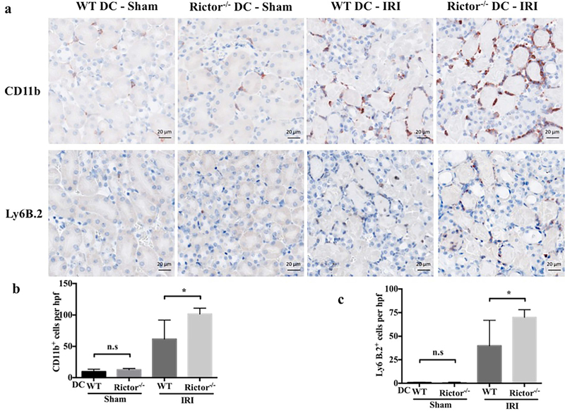
Figure 7|. Inflammatory cell infiltration is enhanced significantly in Rictor−/− DC kidneys following renal IRI.
Kidney sections from sham-operated and post-IRI WT and Rictor−/− DC mice were stained by immunohistochemistry for CD11b and Ly6B.2. (a) Representative micrographs and (b) quantitative analysis of cells/hpf are shown. Images are 400× magnification; scale bars represent 20 m. Data are means +1SD from n=4–6 mice/ per group; *P < 0.05; ns = not significant.

