. 2018 Sep 25;11(3):373–376. doi: 10.3400/avd.cr.18-00061

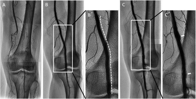
Fig. 1 (A) Angiogram showing total occlusive lesion in the left distal superficial femoral artery to proximal popliteal artery. (B/B′) A 6.0×120 mm Zilver PTX stent was implanted in the occlusive lesion (white dotted line). The stent size was selected to match the vessel size. (C/C′) Angiogram showing peri-stent staining (white arrows) at 21 months after Zilver PTX implantation.

Fig. 1 (A) Angiogram showing total occlusive lesion in the left distal superficial femoral artery to proximal popliteal artery. (B/B′) A 6.0×120 mm Zilver PTX stent was implanted in the occlusive lesion (white dotted line). The stent size was selected to match the vessel size. (C/C′) Angiogram showing peri-stent staining (white arrows) at 21 months after Zilver PTX implantation.