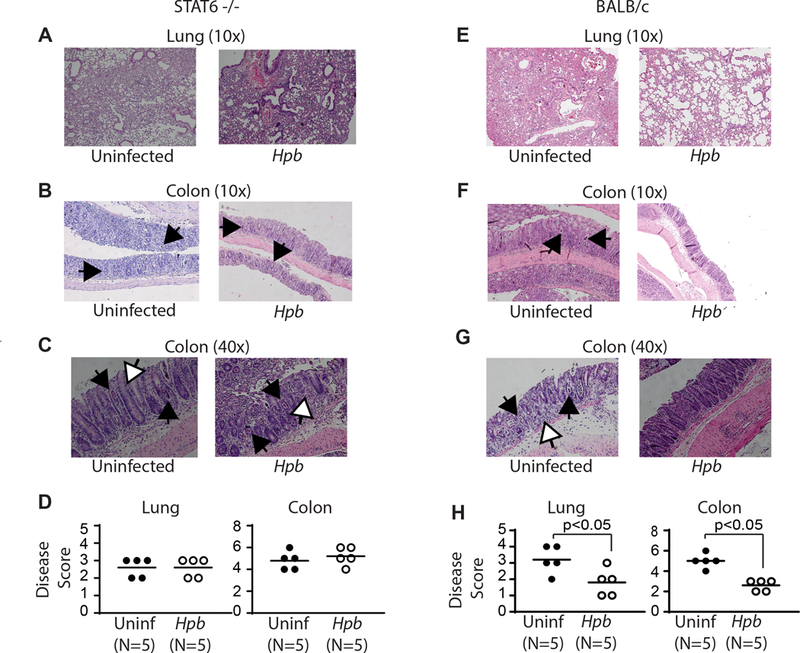
Figure 2. Helminthic suppression of GVHD-related organ damage requires recipient STAT6.

Histopathology of lung (A) and colonic (B-C) tissues from uninfected and Hpb-infected STAT6–/– or WT BALB/c BMT mice six days after BMT. Histopathological analysis was performed in 6-μm-thick, hematoxylin-and-eosin-stained samples. (D) Severity of inflammation in lung and colon, with scoring as described in Materials and Methods. GVHD-related colitis was characterized by mononuclear cell infiltrates, apoptotic cells filling crypts (black arrows) and apoptotic bodies (white arrows). Dot plot distribution with mean of cumulative data from multiple samples. Each dot represents a single sample and the bars the mean values (N=number of samples; p: NS between uninfected (Uninf; black filled dots) and Hpb-infected (white empty dots) STAT6−/− recipients; p<0.05 between uninfected (Uninf; black filled dots) and Hpb-infected (white empty dots) WT BALB/c recipients).
