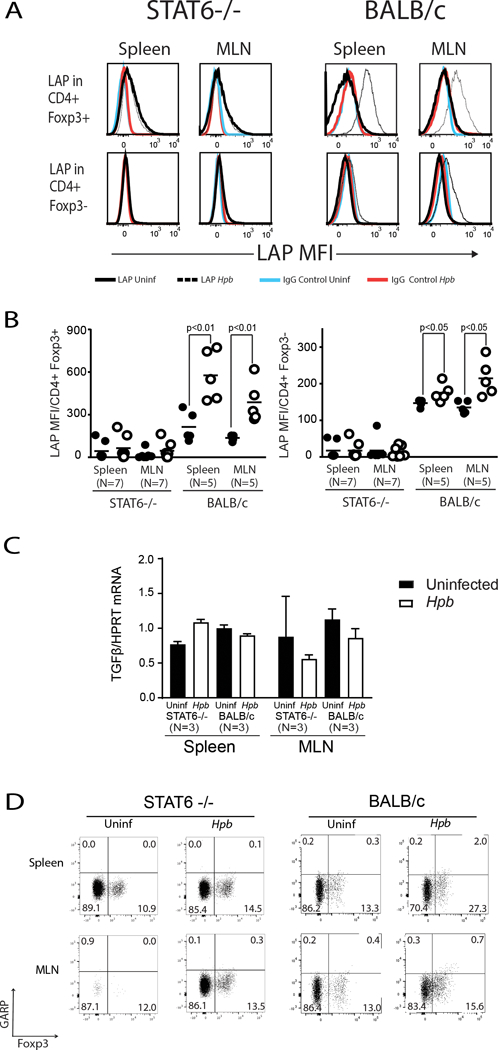
Figure 5. Helminth-induced expression of pro-TGFβ requires STAT6.

Freshly isolated splenic and MLN cells from Hpb-infected and uninfected 8–9 week-old male STAT6–/– or WT (BALB/c) mice were stained for CD3, CD4, Foxp3 and latency-associated peptide (LAP) or CD3, CD4, Foxp3 and control IgG. (A) Histograms showing mean fluorescence intensity (MFI) of LAP or control IgG staining on Foxp3+ CD4 or Foxp3- CD4 T cells from Hpb-infected (white empty dots) or uninfected (black filled dots) mice. (B) Dot plots representing cumulative data from experiments as in A, showing mean±SD for LAP staining from several independent determinations (N: the number of independent determinations; p values as indicated on the figure). (C) TGFβ mRNA content in purified splenic or MLN CD4 T cells from uninfected (Black rectangles) or helminth-infected (white rectangles) STAT6−/− or WT BALB/c mice were displayed as the ratio of TGFβ mRNA to HPRT mRNA. (N: number of independent measurements; p: not significant between uninfected and helminth-infected groups). (D) Spleen and MLN cells from uninfected and helminth-infected STAT6−/− or WT BALB/c mice were stained for CD3, CD4, Foxp3 and GARP. Cells were gated on CD3+ CD4+ T cells. Numbers represent the percentage of events in each quadrant. Data are representative example from at least two samples for each set.
